
Magnetic resonance imaging is a highly accurate way to diagnose internal organ pathologies. The method is due to its informative, noninvasive and painless nature. The absence of radiation exposure allows the technique to be used several times – without limitations. Today we will consider MRI of the knee joint – what it is, how the procedure is performed, how safe it is.
The essence of the method
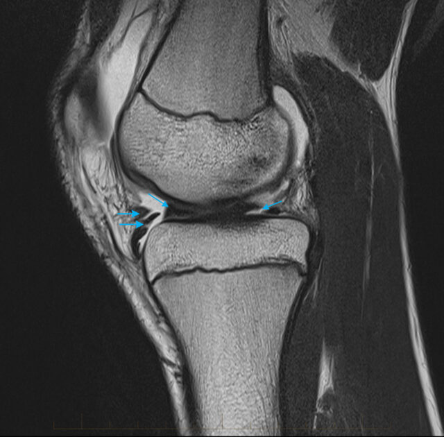
MRI of the knee joint is a diagnostic method based on nuclear magnetic resonance. The joint is affected by the strongest magnetic field. The response of the device is the manifestation of electromagnetic waves, which are processed by a special computer program, and converted into a high-precision image.
The MRI of the knee joint is used to diagnose pathologies of ligaments, tendons, cartilage, meniscus and bone tissue. By studying the results, the specialist identifies abnormalities of the entire joint or individual defects of the studied area – cracks, bruises, damage. The information obtained during the procedure allows you to make a diagnosis, assess the effectiveness of treatment, to determine the need for surgical intervention.
With proper preparation of the patient for the session and coordination of all contraindications, tomography is completely safe. The application of a magnetic field has no effect on the human body. Contrast agent does not pose any danger to the patient either, if there is no allergic reaction to the active substance.
Advantages of MRI of the knee joint:
- high informativeness;
- painlessness and noninvasiveness;
- obtaining high-quality images;
- absence of radiation exposure;
- technological safety;
- minimal contraindications;
- No special preparation and recovery period.
The method has no disadvantages, only a high cost. But this is all relative when it comes to the health and life of the patient.
In what diseases is carried out
Behavioral MRI of the knee joint is prescribed by a traumatologist, orthopedist, oncologist. Read more here mricfl.com.The reason may be to clarify the diagnosis or to check the effectiveness of therapy.
The main pathologies of the musculoskeletal system in the knee area:
- Osteoporosis of the knee joint – bone fragility due to a lack of calcium.
- Gonarthrosis – a degenerative lesion of the knee joint.
- Damage to ligaments, tendons, meniscus of the knee.
- Lyme disease is an infection transmitted by ticks. It is caused by osteoporosis of an unusual course.
- Baker’s cyst – herniation in the area of the knee.
- Osteochondropathy – aseptic necrosis of the cancellous bone.
- Rheumatoid arthritis – loss of mobility in the joints.
- Osteomyelitis – a purulent inflammatory process affecting all elements of the bone.
- Dysplasia – abnormal formation of the knee joints.
- Marfan syndrome – increased stretching of connective tissue.
- Inflammations of the knee joint.
Tomography is prescribed for injuries of different nature, infection of the knee, suspicion of neoplasm development, and treatment monitoring.
Indications for diagnosis

Pathologies of the knee joint are most often manifested by pain in this area. Severe stages of the disease are characterized by limited mobility. The knee is an important element of daily life. Therefore, you should consult a qualified specialist at the first symptoms.
Indications for an MRI scan of the knee joint:
- Persistent pain in the kneecap;
- Complete or partial decrease in mobility;
- swelling of the knee – excessive fluid in the intercellular space;
- deformation of the joint – appearance of hematomas, bruises, redness;
- affection of soft tissues around the knee area;
- internal bone fractures not visible on X-rays;
- sports tears and sprains;
- small clicking sounds when walking.
The presence of these symptoms indicates pathology in the patient’s body. It is better to undergo diagnostics in time to identify the degree, complexity of the disease and prescribe adequate therapy.
Preparation for the procedure
Before the examination, you need to tell the doctor about all contraindications, if they are not reflected in the outpatient card. Patients with kidney and urogenital system diseases should undergo tests. The fact is that the contrasting substance is difficult to eliminate from the body and sometimes causes allergic reactions. It is also better to test for allergens a couple of days before the examination.
Preparing for an MRI of the knee joint does not require any special diets. But with the introduction of a contrasting substance, a few hours before the procedure, it is necessary to completely refuse to eat, because the drug can provoke nausea and vomiting. You should remove all jewelry and metal objects. Make sure that there are no zippers, rivets or aluminum buttons on your clothes that you will wear during the session. It is better to use a disposable cape offered at the clinic.
How the procedure is conducted
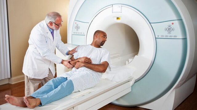
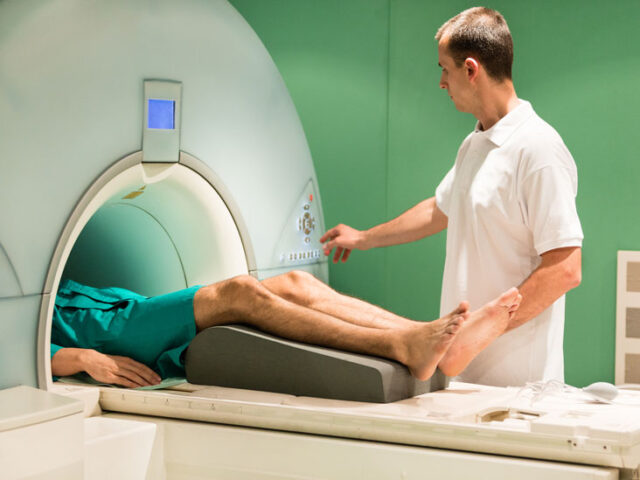
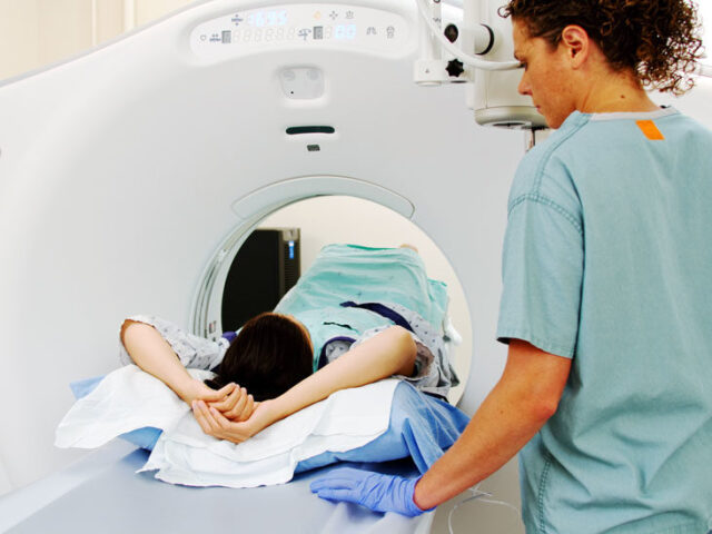
The patient lies down on a special retractable medical table. The patient’s arms and legs are fixed with straps so that there is no mobility during the scan. Next, the patient is put on headphones and the table is slid into a special bulb where the imaging process will take place. If a procedure with the introduction of a contrasting substance is indicated, an intravenous puncture is given and an IV is connected beforehand.
The specialist goes into another room, from where he or she observes the scanning process. There is two-way communication between the doctor and the patient, so the doctor will come to your aid at any minute. If you are not claustrophobic, the procedure should not cause much emotion. If you are still a little scared, ask the doctor for a sedative. If you follow the diagnostic rules, the CT scan is fast and safe.
The duration of a standard MRI of the knee joint is 30 minutes, with contrast – 50 minutes. The description is issued on the same day, but it takes a certain amount of time for the specialist to do so.
Results
Description of the conclusion is – 30-60 minutes. The patient is given a disk with recording, high-resolution image, information on paper. The result can be picked up at the clinic, or it can be sent to an email address. The signal that the description is ready, the patient receives an SMS-message or the patient is invited personally. It all depends on the clinic where you are being examined.


